Frequently Asked Questions: Hydrocolloid Dressings
Contents
- 1 Introduction
- 2 What are hydrocolloid dressings?
- 3 What are the main indications for hydrocolloid dressings?
- 4 Are there any side effects of hydrocolloid dressings?
- 5 How much fluid can hydrocolloid dressings absorb?
- 6 What is the role of hydrocolloid dressings in maggot therapy?
- 7 What is the role of hydrocolloids in hypertrophic scars and keloids?
- 8 Have hydrocolloids been rendered obsolete by newer dressing types?
- 9 Are hydrocolloid dressings cost effective?
- 10 Do hydrocolloid dressings reduce pain?
- 11 Is there any difference between brands?
- 12 Is colour duplex imaging possible through hydrocolloid dressings?
- 13 How effective are hydrocolloid dressings for partial thickness burns?
- 14 Are hydrocolloid dressings contraindicated in diabetes?
- 15 What are the effects of a hydrocolloid dressing on bacterial growth?
Introduction
Hydrocolloids are among the most widely used modern dressings; but that does not necessarily mean that they are widely understood.
This article aims to provide answers to many of the questions that users might ask. It is not intended to be the final word; rather the opposite. These answers are written to be a starting point and no more. Like every article in World Wide Wounds, it can be amended or extended following readers’ suggestions and additions.
What are hydrocolloid dressings?
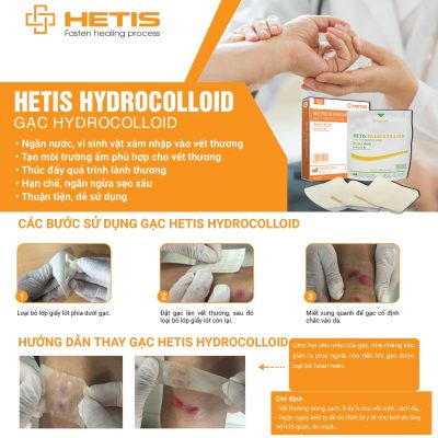
Hydrocolloids are a type of dressing containing gel-forming agents, such as sodium carboxymethylcellulose (NaCMC) and gelatin. In many products, these are combined with elastomers and adhesives and applied to a carrier – usually polyurethane foam or film, to form an absorbent, self adhesive, waterproof wafer.
In the presence of wound exudate, hydrocolloids absorb liquid and form a gel, the properties of which are determined by the nature of the formulation. Some dressings form a cohesive gel, which is largely contained within the adhesive matrix; others form more mobile, less viscous gels which are not retained within the dressing structure.
In the intact state most hydrocolloids are impermeable to water vapour, but as the gelling process takes place, the dressing becomes progressively more permeable. The loss of water through the dressing in this way enhances the ability of the product to cope with exudate production [1].
One feature of hydrocolloids that is appreciated by clinicians is wet tack; unlike most dressings, they can adhere to a moist site as well as a dry one.
Reference 1: Thomas S., Loveless, P. A comparative study of the properties of twelve hydrocolloid dressings. World Wide Wounds, July 1997; [Full Text: /1997/july/Thomas-Hydronet/hydronet.html]
What are the main indications for hydrocolloid dressings?
Hydrocolloids are easy to use, require changing only every 3-5 days, and do not cause trauma on removal. This makes them useful for clean, granulating, superficial wounds, with low to medium exudate.
Hydrocolloids provide effective occlusion; with dry wounds, they can have a softening effect, and they have been used to prevent the spread of MRSA (by providing a physical occlusive barrier).
Reference: Thomas, S., Hydrocolloids Journal of Wound Care 1992:1;2, 27-30
Are there any side effects of hydrocolloid dressings?
Contact dermatitis
Hydrocolloid wound dressings have been in use for some 20 years, and have rarely been associated with allergic contact dermatitis. However, some hydrocolloid dressings contain the pentaerythritol ester of hydrogenated rosin as a tackifying agent, and this substance retains the sensitizing potential of colophony.
Reference: Sasseville D, Tennstedt D, Lachapelle JM: Allergic contact dermatitis from hydrocolloid dressings. Am J Contact Dermat 1997 Dec;8(4):236-238
How much fluid can hydrocolloid dressings absorb?
The ability of hydrocolloids to absorb fluids varies considerably over time, and between products. Laboratory studies [1] suggest that the dressings may not be suitable for medium to high exuding wounds. Other research [2] suggested that when properly applied, the dressings might reduce the amount of exudate.
Reference 1: Thomas S., Loveless, P. A comparative study of the properties of twelve hydrocolloid dressings. World Wide Wounds, July 1997; [Full Text: /1997/july/Thomas-Hydronet/hydronet.html]
Reference 2: Thomas S., Fear M., Humphreys J., Disley L., Waring MJ. The effect of dressings on the production of exudate from venous leg ulcers. WOUNDS 1996;8(5):145-150
What is the role of hydrocolloid dressings in maggot therapy?
Despite decades of experience in Maggot therapy, selecting appropriate dressing materials continues to be a problem. The dressing has to (1) prevent the maggots from escaping, (2) permit oxygen to reach the maggots, (3) facilitate drainage, (4) allow inspection of the wound, (5) require minimal maintenance, and (6) be of low cost.
One centre developed a two-layered cagelike dressing, the bottom layer of which comprised a hydrocolloid pad, applied to the surrounding healthy skin and covered by a fine chiffon or nylon mesh. Liquefied necrotic tissue drained through the mesh and was absorbed in a top layer of gauze, which was replaced periodically. Thus it was possible to contain the maggots within the wound by means of readily available materials.
Reference: Sherman R. A., A new dressing design for use with maggot therapy. Plast Reconstr Surg 1997 Aug;100(2):451-456
What is the role of hydrocolloids in hypertrophic scars and keloids?
Silicone gel sheeting has been investigated for use in the treatments of keloids and hypertrophic scars. Its mechanism of action may be related to scar hydration. One randomized controlled prospective study set out to evaluate a hydrocolloid occlusive dressing that also acts by promoting a moist environment. Scar size and volume, color, patient symptoms, and transcutaneous oxygen measurements were taken.
The study found significantly reduced itching, reduced pain and increased pliability for both treatments, used over two months. The authors concluded that hydration of the scar offered symptomatic improvement, but no change in physical parameters.
Reference: Phillips T. J., Gerstein A. D., Lordan V., A randomized controlled trial of hydrocolloid dressing in the treatment of hypertrophic scars and keloids. Dermatol Surg 1996 Sep;22(9):775-778
Have hydrocolloids been rendered obsolete by newer dressing types?
Over recent years, many new dressings have appeared on the market, but few new dressing types. The continuing success of hydrocolloids depends largely on their effectiveness as occlusive dressings. Any new dressing has to match or better their performance and/or compete on price. Currently, manufacturers of polyurethane foam dressings are promoting them as an alternative to hydrocolloids.
Few studies compare hydrocolloids with newer dressing types. In one randomised controlled clinical study involving 100 patients with leg ulcers and 99 patients with pressure sores in the community, a ‘hydropolymer’ dressing and a hydrocolloid dressing were compared. Statistically significant differences in favour of the hydropolymer dressing were detected for dressing leakage and odour production, but no statistically significant differences were recorded in the number of patients with either leg ulcers or pressure sores who healed in each treatment group.
The future may see hydrocolloids used more selectively, but they are by no means obsolete.
Reference: Thomas S., Banks V., Bale S., et al. A comparison of two dressings in the management of chronic wounds. J Wound Care 1997 Sep;6(8):383-386
Are hydrocolloid dressings cost effective?
Studies too numerous to cite have established that hydrocolloid dressings are more effective than ‘traditional’ dressings, such as parafin gauze, dry gauze and saline soaks. Despite this, and the relative reduction in cost over the decades, many health professionals continue to use obsolete materials and methods.
When the efficacy of hydrocolloid occlusive dressing technique is compared with conventional wet-to-dry gauze dressing technique, the patient has been shown to benefit with a greater chance of healing, faster healing times, and less pain.
Nursing time is very significantly reduced, because the wound does not need dressing so often (or for so long) dressing time is markedly reduced. Costs are saved in materials alone, before even considering the cost of professional time [1]
Similar results have been found in patients with leg ulcers [2]
Reference 1: Kim Y.C., Shin J.C., Park C.I., et al. Efficacy of hydrocolloid occlusive dressing technique in decubitus ulcer treatment: a comparative study. Yonsei Med J 1996 Jun;37(3):181-185
Reference 2: Ohlsson P., Larsson K., Lindholm C., Moller M.A Comparison of saline-gauze and hydrocolloid treatment in a prospective, randomized study. Scand J Prim Health Care 1994 Dec;12(4):295-299
Do hydrocolloid dressings reduce pain?
Pain is a feature of superficial wounds, such as skin graft donor sites, particularly at dressing changes.
One prospective randomized trial compared parafin gauze and a hydrocolloid dressing, applied on donor sites. The results showed that the hydrocolloid is a less painful dressing than parafin gauze, as well as achieving faster healing of skin graft donor sites [1].
In another study, which involved patients with lacerations, abrasions and minor operation incisions, compared a hydrocolloid dressing with a non-adherent dressing. While time to heal was similar for both groups, patients using the hydrocolloid experienced less pain, required less analgesia and were able to carry out their normal daily activities including bathing or showering without affecting the dressing or the wound. [2]
The precise mechanism involved in the hydrocolloid ability to reduce pain is not fully understood, but some possible explanations have been discussed. [3]
Reference 1: Cadier M. A., Clarke J. A. Dermasorb versus Jelonet in patients with burns skin graft donor sites. J Burn Care Rehabil 1996 May;17(3):246-251
Reference 2: Heffernan A., Martin A. J. A comparison of a modified form of Granuflex (Granuflex Extra Thin) and a conventional dressing in the management of lacerations, abrasions and minor operation wounds in an accident and emergency department. J Accid Emerg Med 1994 Dec;11(4):227-230
Reference 3: Nemeth AJ, Eaglstein WH, Taylor JR, et al. Faster healing and less pain in skin biopsy sites treated with an occlusive dressing. Archives of Dermatology, Vol 127, November 1991, pp 1679- 1683.
Is there any difference between brands?
Yes
There are many diffences in structure, flexibility, dimensions, fluid handling properties and, probably, in other performance parameters. The trouble is, few studies have compared different brands [1]. Because of the shortage of in vitro research, and a complete lack of (published) in vivo research, manufacturers claims tend to be based on indirect comparisons such as comparisons based on rival studies which compared hydrocolloids and parafin gauze. One or two comparisons of ‘patient satisfaction’ have been published, but these have no clinical value. Or indeed any value at all, other than ‘marketing exercises’.
Reference 1 Thomas S., Loveless, P. A comparative study of the properties of twelve hydrocolloid dressings. World Wide Wounds, July 1997; [Full Text: /1997/july/Thomas-Hydronet/hydronet.html]
Is colour duplex imaging possible through hydrocolloid dressings?
Colour flow duplex scanning is an accepted method of determining the patency and haemodynamic status in infrainguinal grafts and native arteries. There are often dressings covering the leg above the vessel to be scanned.
A blinded study compared scanning normal superficial femoral arteries with scans when one of five commonly used dressings were applied to the skin above the artery, in random order. The blinded operator graded the signal produced on a linear analogue scale.
An absorbent material dressing and a bilaminate membrane dressing did not transmit ultra-sound at all. Two thin membrane dressings allowed excellent B-mode and colour flow images, in addition to clear Doppler signals. A thin hydrocolloid allowed a clear B-mode image of each artery to be visualised and an adequate Doppler waveform to be obtained. However colour flow mapping was less than optimal although it was possible in each of the arteries.
In patients who require dressings and who may require colour flow duplex scanning of vessels in the same area, a product that permits ultrasound transmission, thus saving the necessity of removing the dressing for the assessment, clearly has advantages
Reference: Whiteley M. S., Magee T. R., Harris R., Horrocks M., Eur J Vasc Surg 1993 Nov;7(6):713-716
How effective are hydrocolloid dressings for partial thickness burns?
A study compared a hydrocolloid formulation with silver sulphadiazine/chlorhexidine parafin gauze dressings in the outpatient management of small partial skin thickness burns.
Burn wounds were followed until complete re-epithelialization occurred. There were no statistical differences between the groups, with respect to healing time, and patients’ subjective responses to treatment.
However, dressing application (but not removal) was easier in the hydrocolloid group. Furthermore, the that group had significantly fewer dressing changes; a mean of three changes per subject group compared with eight in the silver sulphadiazine/chlorhexidine parafin gauze group. In this study, both modalities were found to be equally suitable and effective for small partial skin thickness burns.
Reference: Afilalo M., Dankoff J., Guttman A., Lloyd J., DuoDERM hydroactive dressing versus silver sulphadiazine/Bactigras in the emergency treatment of partial skin thickness burns. Burns 1992 Aug;18(4):313-316
Are hydrocolloid dressings contraindicated in diabetes?
An open randomized controlled study was carried out in 44 patients with diabetes who had necrotic foot ulcers treated with adhesive zinc oxide tape or with an adhesive occlusive hydrocolloid dressing. Fourteen of the 21 patients treated with adhesive zinc oxide tape had their necrotic ulcers improved by at least 50%, compared to six out of 21 with the hydrocolloid dressing (statistically significant). Fifteen patients showed an increase in the area of necrosis during the course of the 5-week study and of these, 10 had been treated with the hydrocolloid dressing. [1]
However, these wounds were necrotic; other clinicians firmly recommend hydrocolloids, particularly for the protection of the wound after the removal of necrotic tissue. [2]
Foot ulcers in people with diabetes, often homogenised by the term diabetic ulcer, usually have both vascular and neuropathic aetiology; it would be unwise to assume that two apparently similar ulcers should be managed the same way. This issue has been controversial since the introduction of hydrocolloids; currently, the best advice would seem to be “use with caution in patients with diabetes.”
Reference 1: Apelqvist J., Larsson J., Stenstrom A., Topical treatment of necrotic foot ulcers in diabetic patients: a comparative trial of DuoDerm and MeZinc. Br J Dermatol 1990 Dec;123(6):787-792 [PubMed abstract] Reference 2: Laing P., Diabetic foot ulcers. Am J Surg 1994 Jan;167(1A):31S-36S [PubMed abstract]
What are the effects of a hydrocolloid dressing on bacterial growth?
Thirty patients with lower limb ulcers of different aetiologies were treated with an occlusive hydrocolloid dressing twice a week for a maximum period of 12 weeks. No antibacterial chemotherapy was utilized. A culture was taken of the exudate of the ulcer before commencement of treatment and weekly or bi-weekly thereafter.
The results showed a mixed flora with prevalence of Staphylococcus aureus. The average duration of the treatment period was 67 days. The average interval between dressing changes was 4.1 days. Subsequent bacterial cultures showed a persistence of the original flora, but there was no correlation between the type of flora present and clinical evidence of infection or between the type of flora present and the rate of healing of the ulcer [1].
In another study, the bacterial flora of chronic venous ulcers treated with an occlusive hydrocolloid dressing were studied over eight weeks. The flora was generally stable. Once a species was present, it remained with the exception of Pseudomonas, which appeared to be inhibited by the dressing. Twelve out of 20 ulcers contained anaerobic bacteria and healing did not appear to be impaired by the presence of any particular species of bacteria [2].
Reference 1: Annoni F., Rosina M., Chiurazzi D., Ceva M., The effects of a hydrocolloid dressing on bacterial growth and the healing process of leg ulcers. Int Angiol 1989 Oct;8(4):224-228
Reference 2: Gilchrist B., Reed C., The bacteriology of chronic venous ulcers treated with occlusive hydrocolloid dressings. Br J Dermatol 1989 Sep;121(3):337-344


